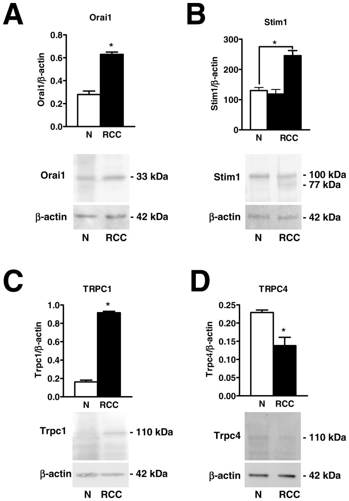
Figure 4. Orai1, Stim1, and TRPC1 proteins are over-expressed in endothelial progenitor cells isolated from patients suffering from renal cellular carcinoma.
Western blot and densitometry depicting the significant elevation in Orai1 (A), Stim1 (B), and TRPC1 (C) proteins in RCC-EPCs as compared to N-EPCs. Conversely, TRPC4 protein (D) is down-regulated in RCC-EPCs. Blots for Orai1, Stim1, TRPC1 and TRPC4 representative of 4 different experiments are shown in the lower panel. Lanes were loaded with 20 μg of proteins. Major bands of the expected molecular weight were observed in both cell types. One additional band of 77 kDa was detected by anti-Stim1 in RCC-EPCs. When both Stim1 bands (77 and 100 kDa) were compared to the single band detected at 100 kDa in N-ECFCs, the expression of Stim1 protein became significantly higher in RCC-EPCs (see text for further explanations). Each bar in the upper panel represents the mean±SE of the densitometric analysis of four different experiments. The asterisk indicates p<0.01 (Student's t-test).

