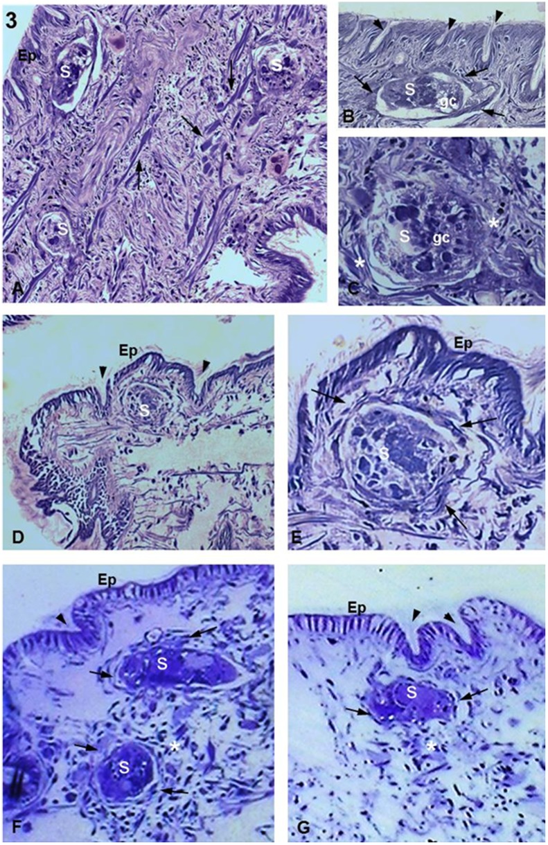
Figure 3. Biomphalaria tenagophila Cabo Frio strain infected with S. mansoni.
(A) 1 h after infection. Three sporocysts (S) are seen sectioned in distinct planes, with one appearing close to the epithelial surface (asterisk). There is an absence of a tissue response or cellular infiltration. Arrows indicate muscle fibers. Magnification 20X. (B,C) 1 h after infection. Details of an S inside the snail tissue showing the germinal cells characteristic of the sporocyst (germ cells; gc). In (B), there is a thin layer of fibrous cells already surrounding the parasite (arrows). By contrast, (C) shows fibrous cells (asterisks) that are not forming a layer around the parasite. Arrowheads indicate crypts. Magnifications 60X. (D) 5 h after infection. Low magnification showing an S located below the tegument of the snail and between two crypts (arrowheads). There is no visible damage on the epithelial surface (Ep). Magnification 20X. (E) 5 h after infection. Details of an S near to the Ep showing a small concentration of fibrous cells (arrows) around the sporocyst forming a thin layer. Magnification 63X. (F,G) 10 h after infection showing two and one S, respectively, close to the Ep with no damage or signs of parasite invasion. The images also show parasites close to the epithelial crypts (arrowheads) and an unattached concentration of fibrous cells (arrows) forming thin layers around the parasites. There is a large concentration of fibrous cells in (F) comparing to (G) (asterisks). Magnification 63X.

