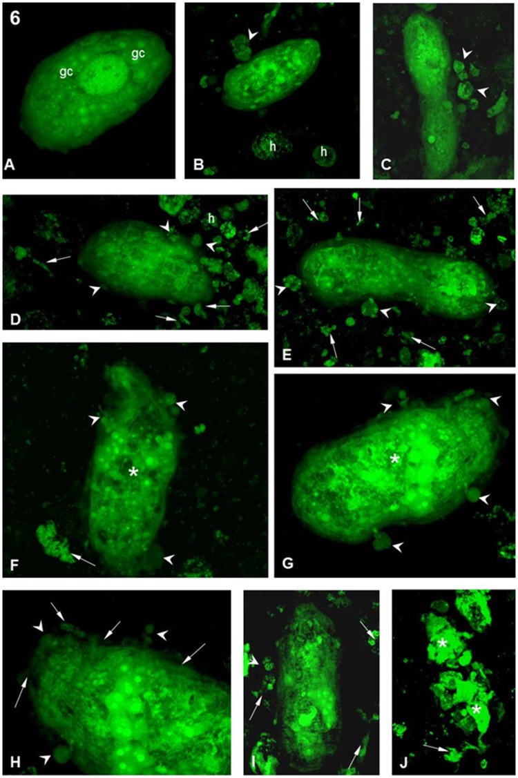
Figure 6. Interaction between hemocytes from the B. tenagophila Taim strain and S. mansoni sporocysts.
(A) After 1 h of interaction, showing a sporocyst with normal morphology and no attached hemocyte. Various germinal cells are present (white arrows). Magnification 40X. (B,C) After 1 h of interaction, showing sporocysts with a few attached hemocytes (arrowheads). ‘h’ indicates free hemocytes. Magnification 40X. (D,E) After 5 h of interaction, showing porocysts with several attached (arrowheads) and free h. There is also a great quantity of cellular debris (arrows). Magnification 40X. (F,G) After 10 h of interaction, several attached hemocytes (arrowheads) are seen on the sporocyst. The parasite morphology is showing disturbance of the internal organelles (asterisks). Cellular debris can also be seen (arrows). Magnification 40X. (H) After 10 h of interaction detailing the anterior region of the sporocyst. There is a hemocyte adhered to the surface (arrowheads) and the disarrangement of the syncytial layer on the surface of the parasite. Magnification 63X. (I,J) After 10 h of interaction. sporocysts with a few attached hemocytes (arrowheads) can be seen, as well as a large amount of cellular debris around the parasite (arrows). The sporocyst in (J) is completely destroyed and is now in several parts (asterisks). Magnification 40X.

