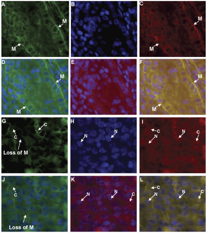
Figure 4. Fluorescence immunostaining with anti-EpEx and anti-Ep-ICD antibodies in aggressive and non-aggressive papillary thyroid carcinomas.
Secondary antibodies are FITC-anti-mouse (green) and TRITC-anti-rabbit (red). A-F images from a non-aggressive PTC; G-L Images from an aggressive PTC. A,G) EpEx; B,H) DAPI; C, I) Ep-ICD; D) EpEx and DAPI (A & C merged); E) Ep-ICD and DAPI (B & C merged); F) EpEx, Ep-ICD, and DAPI (A, B, C merged). J) EpEx and DAPI (G & I merged); K) Ep-ICD and DAPI (H & I merged); L) EpEx, Ep-ICD, and DAPI (G, H, I merged). M, Membranous staining; C, Cytoplasm staining; N, Nuclear staining. Original magnification × 400.

