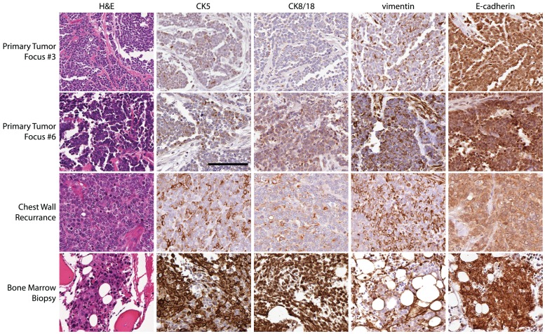
Figure 1. Human breast cancer specimen displays morphological and biochemical evidence suggestive of EMT and MET.
Images of the patient’s primary tumor focus #3 (top row), focus #6 (second row), chest wall recurrence (third row), and bone marrow biopsy (bottom row) showing H&E staining and immunohistochemistry for CK5, CK8/18, vimentin, and E-cadherin.

