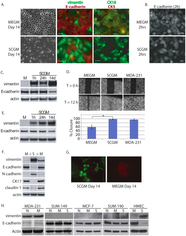
Figure 4. Evidence for morphologic and phenotypic changes consistent with in vitro EMT.
A. Phase contrast or immunofluorescence images of DKAT cells cultured in either MEGM (upper) or SCGM for 14 days (lower). Phase contrast images were acquired with a 20 × objective, scale bar = 40 µm. IF images were acquired with a 40 × objective, scale bar = 20 µm. Cells were stained simultaneously for vimentin (green) and E-cadherin (red), or CK18 (green) and CK5 (red). B. E-cadherin immunofluorescence images of DKAT cells cultured in MEGM or SCGM for 2 hours. C. Total cell lysates from DKAT cells cultured in MEGM or SCGM for 1 h, 24 h, or 14 days were analyzed by western blot for vimentin, E-cadherin and actin. D. Scratch wound healing assay comparing DKAT cells grown in MEGM or SCGM (14 days) and MDA-MB-231 cells. Images (upper panel) were taken at the time of the scratch (T = 0) and 12 hours later with a 5 × objective. Scale bar = 200 µm. Results (lower panel) are expressed as the percentage of the scratch area closed at 12 hrs as measured by ImageJ software, and are an average of 12 scratches per sample. E. Total cell lysates from a clonal DKAT cell line cultured in MEGM or SCGM (14 days) were analyzed by western blot for vimentin, E-cadherin and actin. F. Total cell lysate from passage matched cultures of DKAT cells grown in MEGM, SCGM, or SCGM for 10 passages and then returned to MEGM for 5 passages were analyzed by western blot for epithelial and mesenchymal markers. G. Immunofluorescence (IF) images of subcloned DKAT-SCGM cells cultured in SCGM or MEGM for 14 days. Cells were stained simultaneously for vimentin (green) and E-cadherin (red), and images were acquired with a 40 × objective. H. Total cell lysates from the indicated cell lines cultured in their normal media or switched to MEGM or SCGM for 14 days were analyzed by western blot for vimentin, E-cadherin, and actin expression. N = Normal, M = MEGM, S = SCGM.

