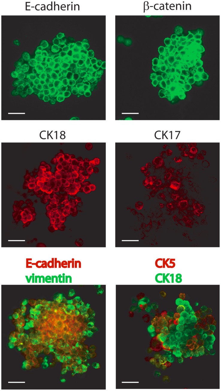
Figure 8. DKAT tumorsphere culture.
Immunofluorescence of DKAT tumorspheres stained for E-cadherin, β-catenin, CK17 and CK18 (upper and middle panels). Tumorspheres were also dual stained for E-cadherin (red) and vimentin (green), or CK5 (red) and CK18 (green) (lower panel). Images were acquired with a 63 × objective. Scale bar = 40 µm.

