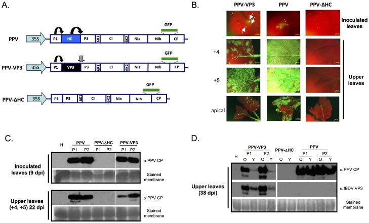
Figure 5. IBDV VP3 is able to functionally replace the HCPro silencing suppressor in a PPV infection.
(A) Schematic representation of full-length cDNA clones derived from PPV and employed to infect N. benthamiana biolistically. The coding sequence of the GFP protein inserted between the NIb and CP cistrons is represented with a green rectangle. Black arrows indicate self-cleavages by the corresponding viral proteases, whereas the grey arrow indicates a cleavage in trans by the action of NIaPro. (B) GFP expression pattern of plants infected with the indicated viruses. Pictures of inoculated leaves collected at 7 days post inoculation (dpi), and the forth (+4) and fifth (+5) leaves above the inoculated one and the most apical leaves collected at 22 dpi were taken under an epifluorescence microscope. (C) Western blot analyses of plant tissue showing GFP foci collected at 9 dpi from inoculated leaves (upper panel) and at 22 dpi from upper non-inoculated leaves (+4 and +5 leaves) (lower panel) of two independent infected plants. (D) Western blot analysis of old (O) and young (Y) leaves collected at 38 dpi from two independent infected N. benthamiana plants. A polyclonal serum specific for PPV CP was used for assessment of virus accumulation, whereas immunoreaction with polyclonal sera specific for IBDV VP3 confirmed the identity of the infecting chimera. The membranes stained with Ponceau red showing the Rubisco are included as loading controls.

