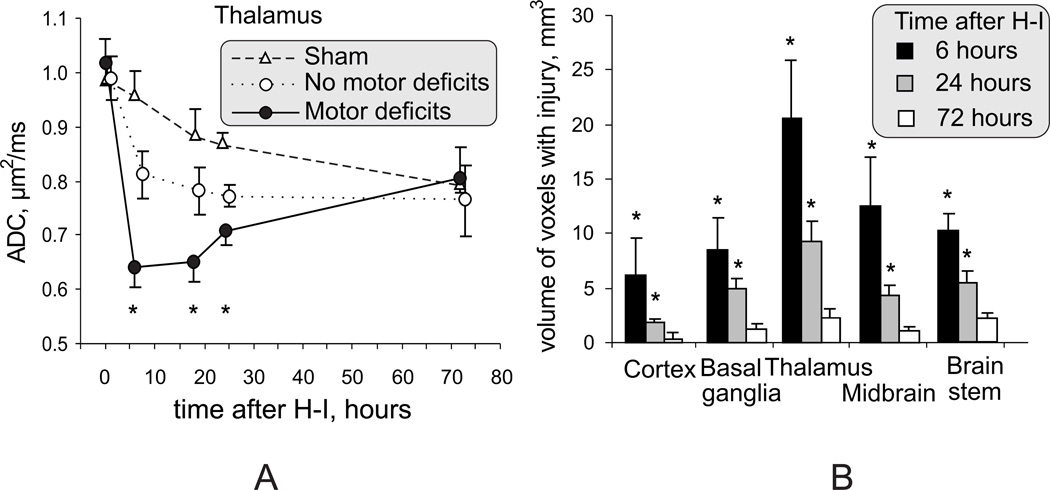
Figure 6.
A. Dynamic changes of ADC in the region of thalamus at the level of third ventricle. Significant ADC decrease persisted in thalamus 6–24 hours after H-I and is an early marker of acute hypoxic thalamic injury. * - p<0.05 comparing motor deficits with group without motor deficits, repeated measures ANOVA.
B. Volume of voxels with ADC below 0.7 µm2/ms in the brains of kits with motor deficits, as an indicator of injury severity, declined from 6 hours to 24 hours after H-I, but remained significant at the 24-hour time point. * - p<0.05, t-test and Bonferroni correction

