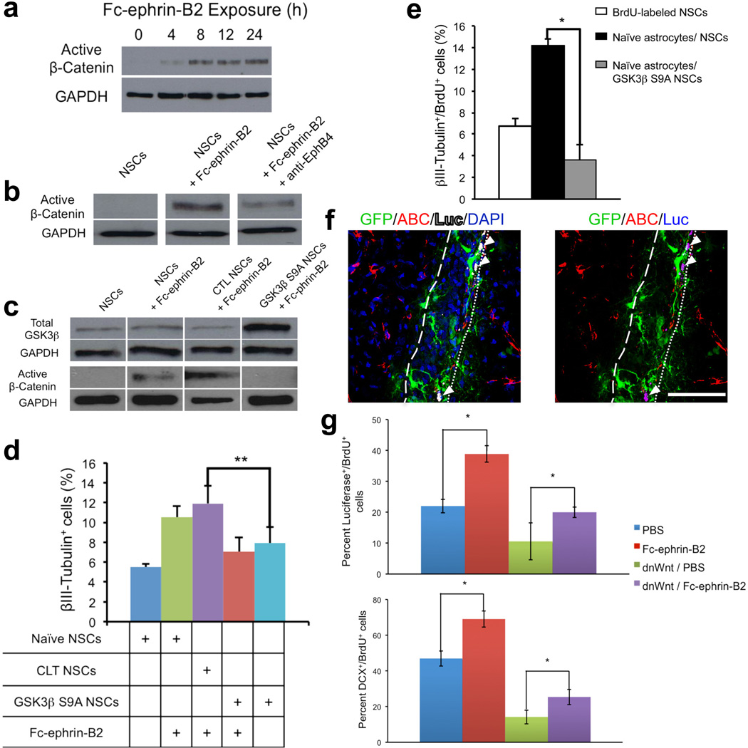
Figure 7.
Ephrin-B2 instructs neuronal differentiation by activating β-catenin independent of Wnt signaling. (a) Fc-ephrin-B2 (10 µg/mL) induced active β-catenin accumulation in NSCs over a 24 hours. (b) However, blocking the EphB4 receptor compromised Fc-ephrin-B2 induction of β-catenin accumulation. (c) NSCs expressing a constitutively active GSK3 β, GSK3 ββS9A, did not accumulate β-catenin in response to Fc-ephrin-B2 (10 µg/mL for 24 hours), in contrast to naïve or empty vector control NSCs (CTL NSCs). (d) Constitutive degradation of β-catenin in GSK3 βS9A NSCs decreased NSCs differentiation into βIII-Tubulin+ neurons in response to Fc-ephrin-B2 (10 µg/mL) vs. empty vector control NPCs (CTL NPCs) (n = 3 experimental replicates). (e) The lack of β-catenin signaling in GSK3 βS9A NSCs also nullified the proneuronal effect of hippocampus-derived, ephrin-B2 expressing astrocytes in co-culture (n=4, experimental repeats). (f) In mice co-infected with Tcf-Luc and dnWnt-IRES-GFP constructs, cells in the SGZ still expressed active β-catenin (ABC) and Luciferase (arrowheads) 24 hours after Fc-ephrin-B2 injection. Scale bar represents 100 µm. (g) In hippocampi co-infected with Tcf-Luc and either dnWnt-IRES-GFP or IRES-GFP construct, then injected with Fc-ephrin-B2 or PBS, Fc-ephrin-B2 increased the percentage of SGZ BrdU+ cells with active β-catenin signaling 24 hours post-injection and the percentage of DCX+/BrdU+ cells by day 5 even with dnWnt present (n=4 brains, 8 sections per brain). Ephrin-B2 thus activates β-catenin signaling and enhances adult neurogenesis independent of Wnt signaling. * indicates P <0.01; ** indicates P <0.05; ± s.d; dotted vs. dashed lines mark SGZ/Hilus vs. GCL/MCL boundaries. Full length blots in Supplementary Fig. 9.

