
Figure 3.
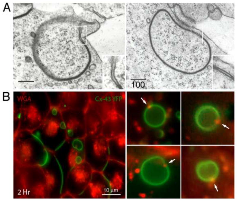
Fine structure and composition of AGJ vesicles. (A) Double-membrane AGJ vesicles can contain a patch of non-junctional membrane where the two membrane layers are separated (enlarged in inserts). These non-junctional membrane patches appear to be derived from plasma membrane that was located immediately adjacent to the GJ plaque and was concomitantly endocytosed. (B) These non-junctional AGJ membrane domains label with extracellularly applied, fluorescence-labeled wheat germ agglutinin (WGA) (red puncta on green AGJ vesicles marked with arrows) in Cx43-YFP expressing cells. Stable Cx43-YFP expressing HeLa cells were incubated for 2–4 h with Alexa594-labelled WGA and examined by fluorescence microscopy. Low magnification survey image (panel 1, left) and high-resolution images of internalized AGJ vesicles (panels 2–5, right) are shown (scale bars = nm in [A], and m in [B]). (Figure 3A is adapted from (Piehl et al. 2007).)