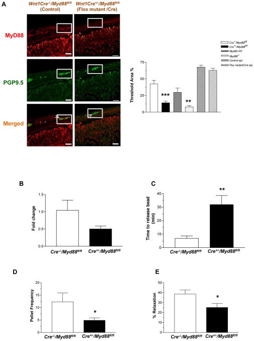
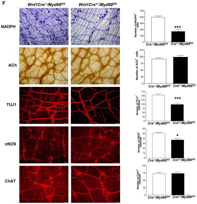
Figure 6. Wnt1Cre+/−/Myd88fl/fl mice have delayed colonic transit and reduced nitrergic neurons.
(A) Wnt1Cre−/−/Myd88fl/fl and Wnt1Cre+/−/Myd88fl/fl mice, and Myd88−/− mice were stained for Myd88 and neuronal marker PGP9.5. Histogram shows percentage of area stained for Myd88 in enteric ganglion or epithelial cells determined by morphometry. (B) Expression of Myd88, by real time-PCR in the enteric ganglia of Wnt1Cre+/−/Myd88fl/fl mice relative to Wnt1Cre−/−/Myd88fl/fl mice. (C) Colonic motility was determined by time to release bead as described in Methods section, and (D) Pellet frequency per hour was assessed in Wnt1Cre−/−/Myd88fl/fl and Wnt1Cre+/−/Myd88fl/fl. (E) Nitrergic relaxation in response to nerve stimulation was assessed in proximal colon of the mice. (F) Representative photographs and histograms of proximal colonic whole mount staining for NADPH Diaphorase, Acetylcholinesterase and terminal ileum whole mount staining for TUJ1, nNOS and ChAT. The number of neurons stained for a specific marker was determined per unit area. Scale bar: 50 μm. Results are mean ± SE. n=6. *P < 0.05, **P < 0.01, ***P < 0.001.


