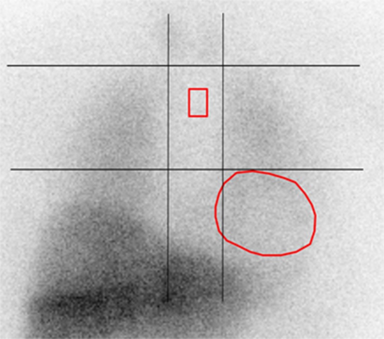
Fig. 1.
The mediastinal ROI and the manually drawn polygonal cardiac ROI on a planar MIBG image. The rectangular mediastinal ROI (size 13 × 20 pixels) was placed in the upper half of the “anatomical landmark square” formed by the lung apexes (upper square border), the upper cardiac border (lower square border) and the medial contours of the lungs (medial square borders). The manually drawn polygonal cardiac ROI was placed over the myocardium including the left ventricular cavity

