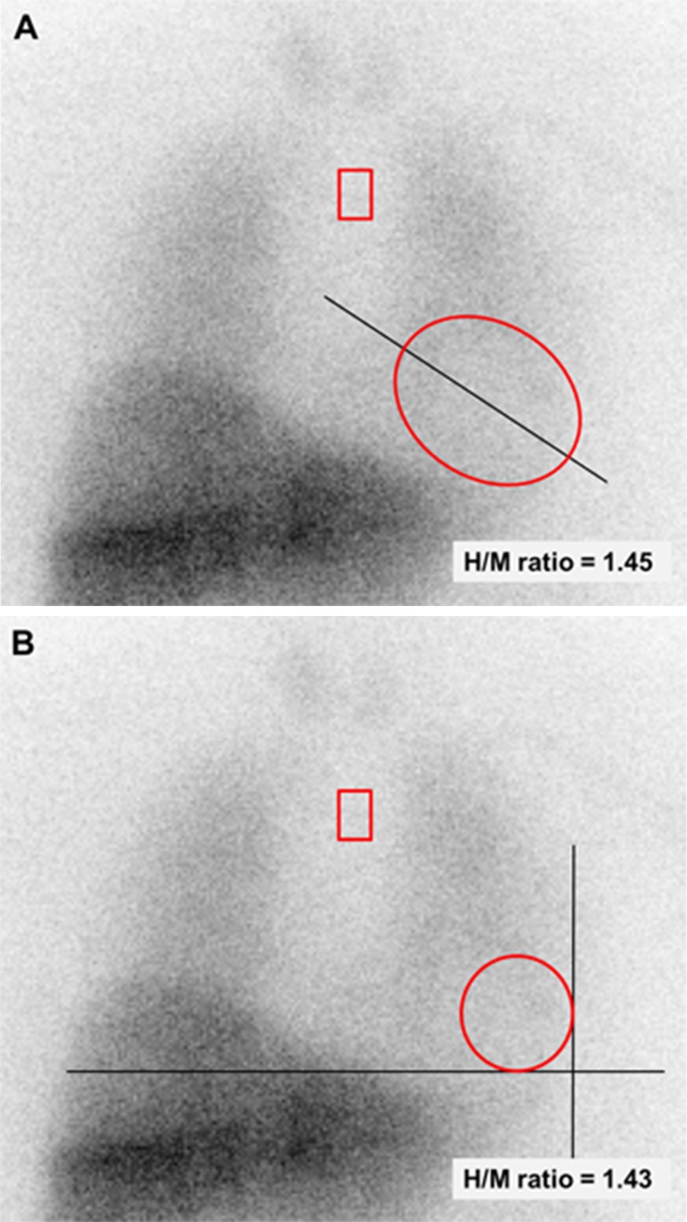
Fig. 2.
Oval and circular fixed-size cardiac ROIs on planar MIBG images. a The oval cardiac ROI was placed over the myocardium including the left ventricular cavity and had a size 60 × 70 pixels (approximately 85 × 100 mm). The long axis of the oval was in line with the heart axis. b The circular cardiac ROI had a predefined radius of 21 pixels (approximately 30 mm) and was placed at the apex of the heart, covering a part of the myocardium and left ventricular cavity. Using the oval and circular cardiac ROIs, the calculated H/M ratios were 1.45 and 1.43, respectively. In this patient the H/M ratio using the polygonal manually drawn cardiac ROI was 1.46

