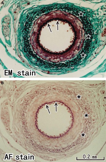
Fig. 1.

Elastic fibers in the arterial wall: a positive control of the present staining methods. The inner elastic lamina (arrows) of a branch of the external carotid artery is clearly identified as a black line in elastica-Masson staining (panel A) and as a bright violet line in aldehyde-fuchsin staining (panel B). The outer lamina was identified as black or violet dots (stars). These panels are prepared at the same magnification: scale bar in panel B.
