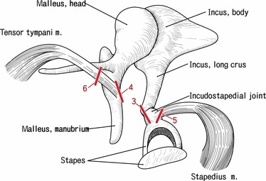
Fig. 2.

A diagram showing the present sectional planes. Right ear ossicles with attachments of the tensor tympani and stapedius muscles are shown, although in the present study, we used both the left and the right sides of the specimens. Bar with 3, 4, 5 or 6 corresponds to the sectional plane shown in the Figs 3, 4, 5 or 6.
