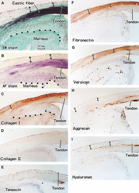
Fig. 4.

Tendon insertion of the tensor tympani muscle to the malleus. A specimen from an 85-year-old man. All panels are adjacent or near sections. (A) Elastica-Masson staining (EM stain). (B) Aldehyde-fuchsin staining (AF stain). (C) Immunohistochemistry (IHC) for type I collagen. (D) IHC for type II collagen. (E) IHC for tenascin-c. (F) IHC for fibronectin. (G) IHC for versican. (H) IHC for aggrecan. (I) IHC for hyaluronan. (C–I) are prepared at the same magnification (scale bars in A–C). Dotted line in (A–C) indicate the tidemark: elastic fibers insert deeply into the uncalcified fibrocartilage. Type I collagen, fibronectin and versican are expressed in the superficial layer of the tendon (double-headed arrow).
