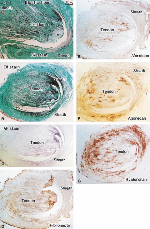
Fig. 6.

Distal parts of the tensor tympani muscle tendon. A specimen from an 85-years-old man (the same specimen as shown in Fig. 4). (A,B) Elastica-Masson staining (EM stain). (C) Aldehyde-fuchsin staining (AF stain). (D) Immunohistochemistry (IHC) for fibronectin. (E) IHC for versican. (F) IHC for aggrecan. (G) IHC for hyaluronan. All panels are prepared at the same magnification (scale bar in A). (A) Muscle–tendon interface of the tensor tympani muscle. (B–G) Adjacent or near sections. At the muscle–tendon interface, elastic fibers converge to a collagenous core (star in A); near the insertion, the fibers gradually occupy almost all cut surface of the tendon (B,C). The tendon sheath also contains abundant elastic fibers. Hyaluronan, versican and fiibronectin display a laminar distribution between the elastic fibers (D–F). Many aggrecan-positive spots are seen in the sheath of the tendon (G).
