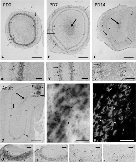
Fig. 2.

Expression and localization patterns of CRMP4 mRNA in the MOB and AOB during postnatal development. (A–D, A′–D′) Representative images of hybridization signals in the MOB on PD0 (A, A′), PD7 (B, B′), and PD14 (C, C′) and in adults (8 weeks old) (D, D′). (A′–D′) Higher magnification of delineated areas in A–D. Arrowheads and arrows indicate the MCL and glomerular layer, respectively. Big arrows in B, C, and D indicate hybridization signals in the subependymal zone. (E,F) Pictures of mirror-image sections of the subependymal zone in the adult OB. In situ hybridization signals of CRMP4 mRNA (E) were colocalized with PSA-NCAM immunoreactivity (F) in the same cells (arrowheads). (G–J) Representative images of hybridization signals in the AOB on PD0 (G), PD7 (H), and PD14 (I) and in adults (J). Arrowheads and arrows in G, H, I, and J indicate the MCL and glomerular layer, respectively. Scale bars: 400 μm (A–D), 100 μm (A′–B′ and G–H), 50 μm (C′,E–F and I–J), 10 μm (D′).
