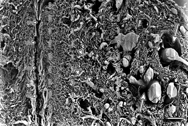
Fig. 1.

Cervix sagittal section – supravaginal part. Vertical interrupted lines divide the vasculature into four zones. The outermost zone is on the far right of the figure. The straight arrows point to the places where perivascular connective tissue was located. The triangles mark the vessels that run transversely and supply the pericanalar vessels. ‘Serpent’ arrows point to the cervical canal. Scale bar: 1000 μm.
