
Fig. 3.
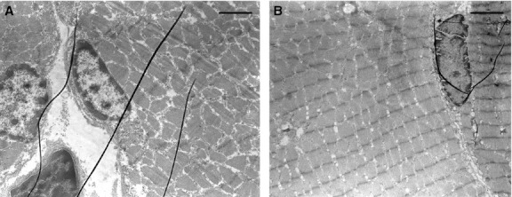
TEM micrographs showing well-preserved muscular fibers with a normal ultrastructure of the nuclei, located peripherally in the zone immediately beneath the cell membrane. Note the different aspect of the serially arranged sarcomeric units in transversal (left) and longitudinal (right) section. Scale bars: 1.7 μm (A); 1.8 μm (B).