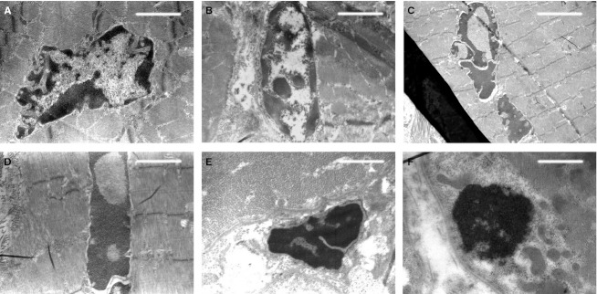
Fig. 4.
TEM micrographs showing, from (A) to (F), muscular fiber nuclei in apoptosis at different progressive stages of chromatin condensation. Note in (E) and (F), with respect to the previous stages, the disruption of the sarcomeric units. Scale bars: 1.5 μm (A, B, D); 2 μm (C); 1 μm (E); 0.75 μm (F).

