
Fig. 1.
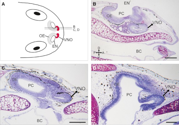
Nasal region in anuran tadpoles. (A) Schematic illustration of the nasal region of a generalised tadpole (dorsal view). Dotted lines (B, C, and D) indicate the estimated planes of the parasagittal sections shown in panels B–D. (B–D) Histological parasagittal sections (stained with cresyl violet) through the nasal region of X. laevis (B, stage N/F54), R. arenarum (C, stage G35), and L. catesbeianus (D, stage G34) showing the relative position of the bean-shaped VNO anteroventral to the olfactory epithelium in the principal chamber (PC). BC, buccal cavity; EN, external naris. Axes indicate: A, anterior; P, posterior; D, dorsal; V, ventral. Scale bar, 200 μm.