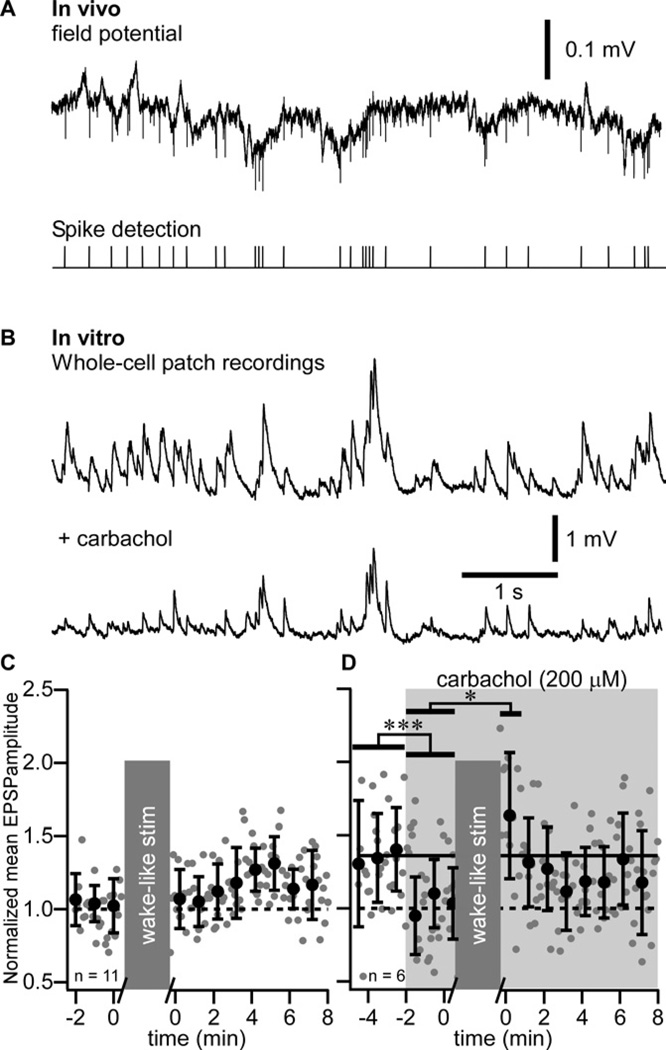
Figure 5. Absence of long-term potentiation after wake pattern of stimulation in vitro.
(A) In vivo field potential recording during waking period of a cat. Extracellular spikes were detected and their timing was used as synaptic stimulation pattern.
(B) In vitro recordings during wake-like pattern of synaptic stimulation in control (Upper trace) and after adding 200 µM of carbachol (Lower trace).
(C) Group data of normalized EPSP amplitude of in vitro whole-cell recordings in control and after wake-like pattern of synaptic stimulation.
(D) Group data of normalized EPSP amplitude of in vitro whole-cell recordings in control and after wake-like pattern of synaptic stimulation in presence of carbachol (shaded area). Gray dots are individual responses amplitude and black circles are the running averages and standard deviation for 12 consecutive responses (1 min). *p<0.05, ***p<0.001, Mann-Whitney test. Grey boxes indicate the 10 minutes stimulation protocol that was used.

