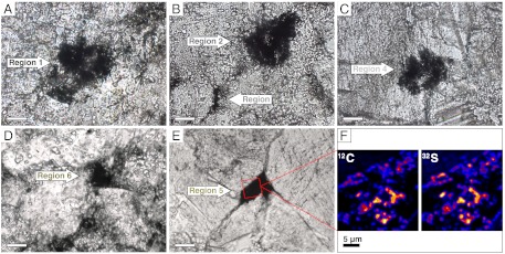
Fig. 2.
Silicified organic matter preserved in the 3.45-Ga SPF stromatolites. (A–E) Transmitted light photomicrographs showing the organic-rich regions analyzed by NanoSIMS. Scale bars, 20 μm. (F) NanoSIMS ion maps of 12C and 34S (red square in E). The coregistration between the abundance of 12C and 34S in the ion images indicates that measured δ34S values derive from sulfur incorporated into organic matter. The organic material is finely dispersed within the chert, forming aggregates that rarely exceed a size of 3 × 3 μm.

