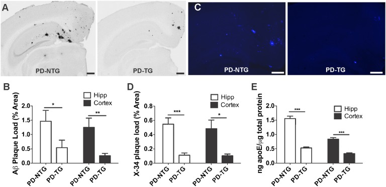
Fig. 2.
LDLR overexpression in PDAPP mice markedly decreases brain Aβ/amyloid deposition and apoE levels. (A) Representative coronal brain sections from 10-mo-old, sex-matched PDAPP+/− mice expressing normal levels of LDLR (PD-NTG), and PDAPP+/− mice overexpressing LDLR (PD-TG). Aβ immunostaining was performed using anti-Aβ antibody (biotinylated 3D6). (Scale bars, 300 μm.) (B) Quantification of the area of the hippocampus or cortex occupied by Aβ immunostaining (n = 9 mice per group). (C) Representative amyloid burden in coronal brain sections from 10-mo-old, sex-matched PD-NTG mice and PD-TG mice. Amyloid was visualized using the congophilic fluorescent dye, X-34. (Scale bars, 100 μm.) (D) Quantification of the area of hippocampus or cortex occupied by X-34 staining (n = 9–10 mice per group). In B and D, groups were compared using the Mann–Whitney U test. *P < 0.05, **P < 0.01, ***P < 0.001. (E) ApoE protein levels measured by sensitive sandwich ELISA in hippocampal and cortical homogenates from PD-NTG and PD-TG mice (at 3–4 mo of age to avoid confounding effects from amyloid plaque deposition; n = 9 mice per group). Differences between groups were assessed using two-tailed Student’s t test (with Welch's correction for E). ***P < 0.001. Values represent means ± SEM.

