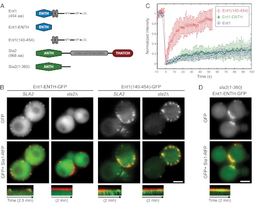
Fig. 2.
The Sla2 ANTH domain is required for stable binding of the Ent1 ENTH domain to the endocytic site. (A) The domain organization of Ent1 and Sla2 and the fragments used in this study. Protein domains and linear motifs for binding to EH-domain proteins (NPF) and clathrin (LIDL) are shown. (B) Localization of Ent1-ENTH-GFP or Ent1(140–454)-GFP together with Sla1-RFP in epsin-depleted cells in the presence or absence of Sla2. GFP and merged images are shown together with kymographs of Ent1 constructs and Sla1-RFP at endocytic patches. (C) FRAP of Ent1-ENTH-GFP and Ent1(140–454)-GFP at endocytic sites in latA-treated epsin-depleted cells. Curves represent the mean ± SD (n = 7–13 separate measurements). The Ent1-GFP FRAP in latA-treated wild-type cells (from Fig. 1C) is shown for comparison. (D) Sla2(1–360) stabilizes Ent1 ENTH at endocytic sites. Localization of Ent1-ENTH-GFP and Sla1-RFP in epsin-depleted sla2(1-360) cells (compare with B). GFP and merged images are shown together with the kymographs. All kymographs are oriented with the cell exterior at the top. (Scale bars: 2 μm in B and D.)

