Fig. 4.
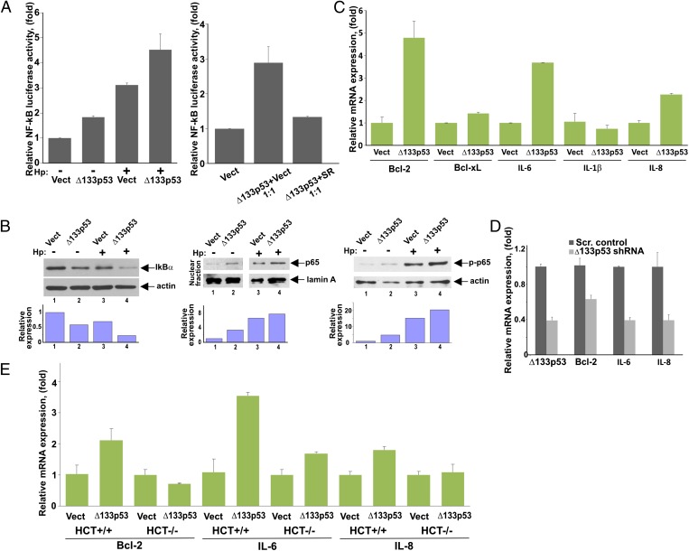
Δ133p53 protein regulates NF-κB activity. (A) NF-κB activity was assessed in SNU-1 cells transfected with either Δ133p53 expression vector (Δ133p53) or empty control vector (Vect) and co-cultured with H. pylori (Hp) (+) for 8 h or left uninfected (−). NF-κB activity was assessed using the pNFκB-Luc reporter. (Right) Reporter analysis was repeated in the presence of the NF-κB inhibitor, IκB SR. SNU-1 cells were cotransfected with Δ133p53 vector and SR (or empty vector) at a 1:1 ratio. (B) SNU-1 cells stably transfected with Δ133p53 or empty control vector were co-cultured with H. pylori for 3 h and analyzed for IκBα protein (Left) as well as nuclear localization of p65 (RelA) protein (Center) and its phosphorylation at serine 536 (Right) by Western blotting. The graphs represent the results of densitometric analysis of immunoblots and depict actin (or lamin A)-normalized protein expression. (C) SNU-1 cells were transfected with either Δ133p53 or empty control vector for 48 h and analyzed for mRNA expression of the indicated NF-κB target genes by qPCR. (D) AGS cells were stably transfected with a specific shRNA against Δ133p53 or scrambled (Scr.) control vector and were analyzed for mRNA expression of Bcl-2, IL-6, and IL-8 by qPCR. (E) Same as in C, but isogenic cell lines HCT116+/+ (WT p53) and HCT116−/− (p53-null) were used.

