Fig. 1.
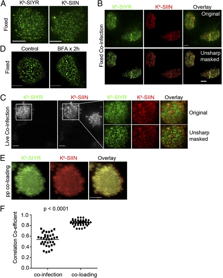
Endogenously generated SIYR– and SIIN–Kb complexes are expressed in distinct cell-surface clusters. (A) (Left) l-Kb cells 4 h p.i. with VV expressing Ub-liberated SIYR were stained with biotinylated 2Cm67, followed by secondary staining with Alexa 488-conjugated Streptavidin. After fixation, cells were imaged with TIRF microscopy; (Right) LKb cells infected with SIIN-expressing VV were fixed, surface stained with 25D1.16 and then stained with an Alexa647-conjugated goat anti-mouse IgG secondary antibody. TIRF was performed using a Leica AF6000× microscope equipped with a 100× objective. (B) Four hours post coinfection with VV expressing Ub-liberated SIYR and SIIN (MOI = 1 for each), l-Kb cells were costained with biotinylated 2Cm67 and Alexa647-conjugated 25D1.16, followed by secondary staining with Dylight488-conjugated Streptavidin. Unsharp mask processing was performed with Metamorph Imaging Series 7.1. (C) Four hours p.i. with VV expressing Ub-liberated SIYR or SIIN (MOI = 1 for each), l-Kb cells were stained live with a mixture of directly conjugated monovalent reagents, Alexa488-conjugated 2Cm67 and Alexa647-conjugated 25D1.16. Stained cells were visualized live with dual-color TIRF. Enlarged images showed the relative localization between Kb–SIYR and Kb–SIIN on the cell surface. Unsharp mask processing was done with Metamorph Imaging Series 7.1. Note that of two cells in field, only one is coinfected, providing an internal specificity control for staining. (D) Kb–SIIN still clustered after abrogation of surface class I MHC delivery by BFA. l-Kb cells were incubated with BFA at 10 μg/mL starting at 4 h p.i. with VV expressing Ub-liberated SIIN (MOI = 1). Two hours later, cells were fixed with paraformaldehyde, surface stained with 25D1.16 and Alexa488-conjugated goat anti-mouse IgG secondary antibody. (E) Exogenous Kb–SIYR and Kb–SIIN complexes colocalize. Human β2-microglobulin (hβ2m)-sensitized l-Kb cells loaded with a mixture of synthetic SIYR and SIIN peptides (5 μM each), stained live with a mixture of Alexa488-2Cm67 and Alexa647-25D1.16 Fab and imaged immediately by dual-color TIRF. (F) Statistical analysis of the colocalization coefficients between exogenous peptide coloading and endogenous coinfection conditions in l-Kb cells. More than 30 images were collected from each condition. After background subtraction, the correlation coefficient R was calculated with the Image Correlation 1o plugin of National Institutes of Health Image J software.

