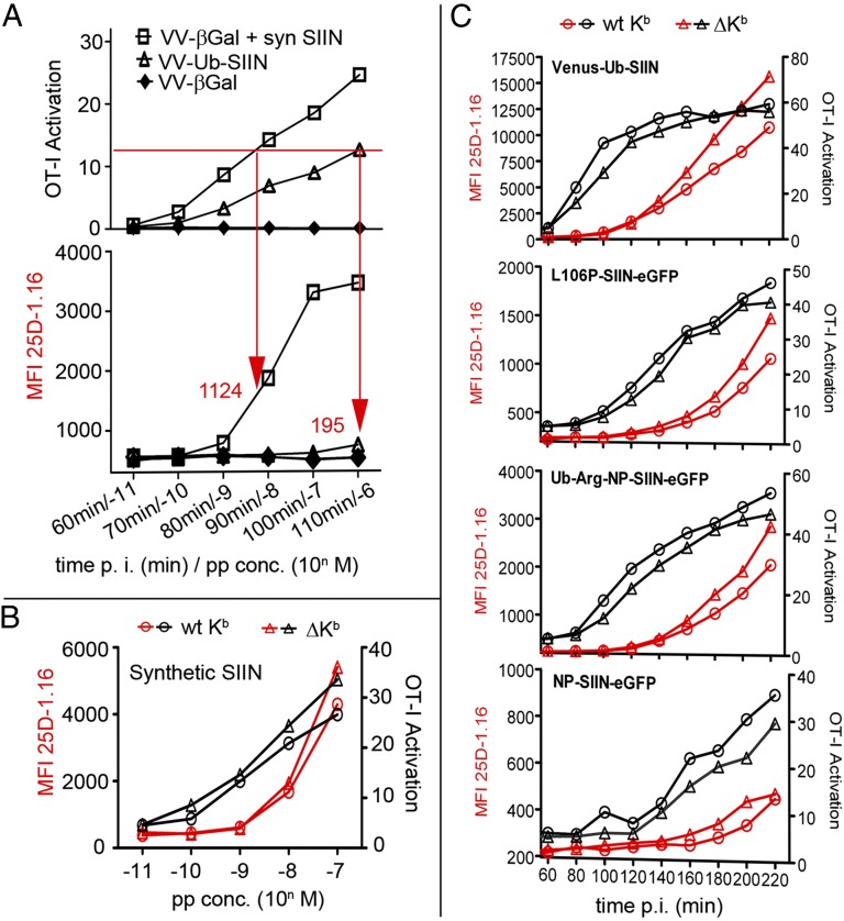
Fig. 5.
Kb–SIIN clustering is associated with increased T-cell sensitivity. (A) L929/Kb cells infected with VV-Ub-SIIN (MOI = 2) or VV-βGal (control) were coincubated with OT1 CD8 T cells (E:T = 1:1) for 1.5 h in the presence of brefeldin A. IFN-γ expression was measured by flow cytometry for intracellular anti-IFN-γ expression, gating for CD8α+ cells. In parallel, L929/Kb cells were stained with 25D1.16 for surface Kb–SIIN expression. For peptide loading, hβ2m sensitized L929/Kb cells were infected with VV-βGal for 2 h followed by peptide incubation for 1 h at 4 °C. Arrows and numbers indicate the MFI of Kb–SIIN required in each condition to achieve equivalent levels of OT1 activation. (B) L929/Kb or tailless L929/ΔKb cells pretreated with human β2m were loaded with SIIN peptide and tested for their ability to activate OT-I cells as in A. (C) OT-I activation tested as in A with L929/Kb or tailless L929/ΔKb cells after infection with indicated recombinant VV expressing SIIN in the form of peptide (Venus-Ub-SIIN) or full-length rapidly degraded (L106P-SIIN-eGFP, Ub-Arg-NP-SIIN-eGFP) or stable (NP-SIIN-eGFP) proteins.

