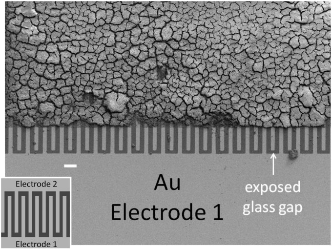
Fig. 1.
SEM of a fully grown WT G. sulfurreducens biofilm grown on a gold interdigitated microelectrode array (IDA). The edges of the array were masked with photoresist, defining the electroactive area where the biofilm grew, which was removed during preparation for SEM imaging. An unmasked edge of the array is shown at the bottom, where alternate microelectrode bands comprising electrode 1 are electrically connected. (Scale bar: 45 μm.) (Inset) Schematic representation of a portion of the IDA depicting 10 of 100 microelectrode bands (not to scale; dimensions provided in the text).

