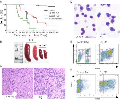
Fig. 1.
Enforced expression of Erg induces development of leukemia in mice. (A) Survival of murine bone marrow chimeras with enforced expression of Erg in hematopoietic cells. Sublethally irradiated mice were transplanted with hematopoietic cells transduced with empty vector control (black line), FLCs transduced with Erg (red line), or post-5FU bone marrow cells transduced with Erg (blue line). Lethally irradiated mice were transplanted with FLCs transduced with Erg (green line). Mice receiving lethal irradiation succumbed to leukemia more rapidly than sublethally irradiated mice. (B) Bone marrow chimeras that had received Erg-transduced hematopoietic cells, and did not develop lymphoid leukemia, developed enlarged spleens (Erg) compared with mice that had received empty vector–transduced cells (Control). (C) The livers of these mice (Erg) displayed heavy infiltration by leukemic cells, compared with healthy control mice (Control). Representative sections taken at 100× magnification are shown. (D) May–Grunwald–Giemsa staining of representative cytocentrifuge preparations of spleen cells is shown. Cytological analysis of spleen cells from leukemic mice (Erg) revealed a predominance of nucleated erythroid cells with basophilic staining of the cytoplasm, compared with control mice (Control). (E) Flow cytometric analysis of erythroid development in the bone marrow. Normal erythroid development was observed in control mice (Control BM). Erythroid development in leukemic mice (Erg BM) was severely perturbed, with a buildup of mCherry+ cells expressing high levels of CD71 and increasing levels of Ter119. (F) A subset of the mCherry+ leukemic erythroblast cells also expressed the megakaryocytic marker CD41 (Erg BM). This was not observed in control mice (Control BM).

