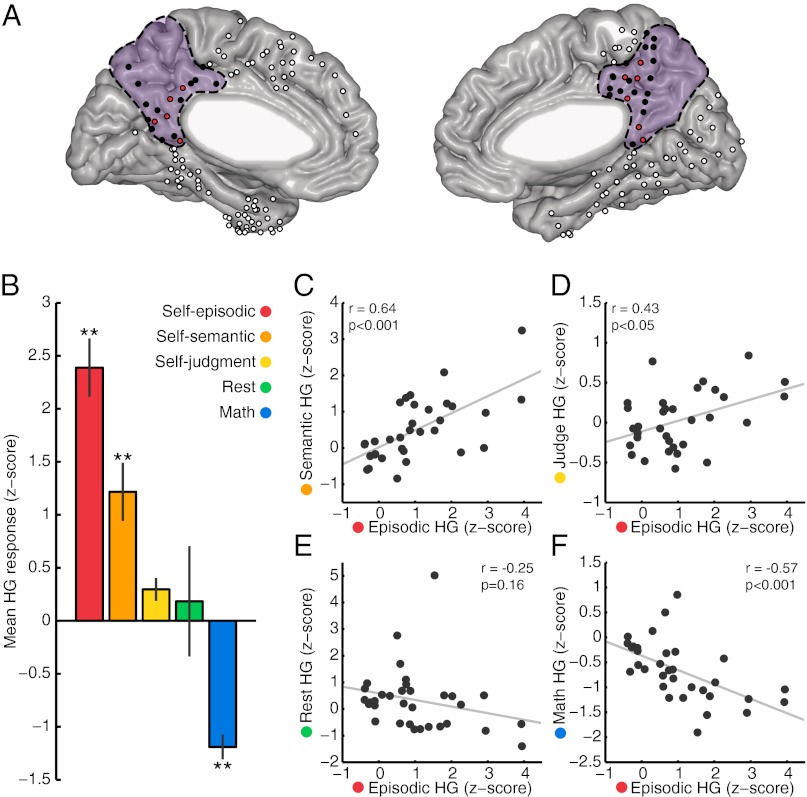
Fig. 2.
Response magnitude across conditions. (A) Electrode locations from all subjects on a standardized Montreal Neurological Institute brain, with the PMC marked in purple. PMC electrodes responsive to the self-episodic condition have red fill and other PMC sites have black fill. Electrodes falling outside the anatomical boundaries of the PMC are filled in white. (B) Mean HG response across conditions for all electrodes that were identified as significantly responsive during the self-episodic condition (11, marked red in A). Mean HG response for these electrodes was significantly different across conditions [one-way ANOVA; F(4,40) = 17.43, P < 0.001], with post hoc test showing self-episodic, self-semantic, and math to be significantly different from all other conditions (**P < 0.01, corrected). Scatter correlation plots of mean HG response for all PMC electrodes (n = 33, marked black in A) comparing the self-episodic condition with self-semantic (C), self-judgment (D), rest (E), and math (F) conditions, respectively.

