Fig. 3.
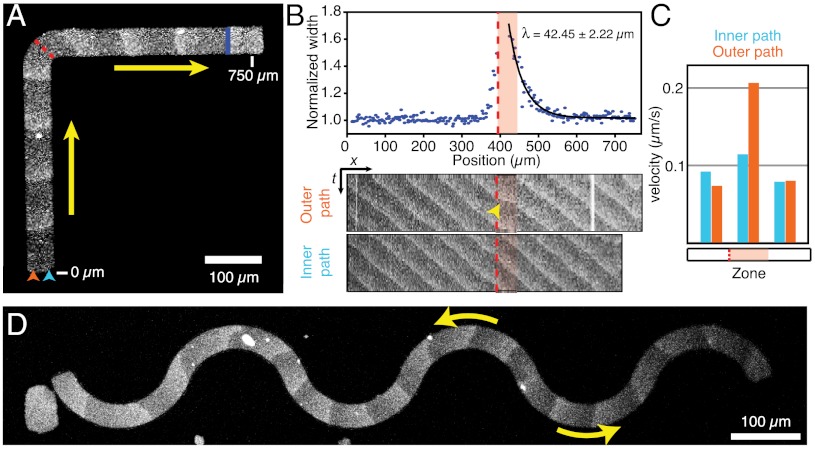
The membrane guides traveling waves. (A) Confocal micrographs of Min protein waves on a L-shaped membrane. Min protein waves turn by 90° following the path of the membrane. (B) Upper: Relative change of protein band width. Exponential fit gives the characteristic travel path length required for realignment of the wave. Lower: Kymographs along the outer (orange arrowhead) or inner (blue arrowhead) path of the membrane shown in A. (C) Velocities within the traveling protein band. During realignment (B, red shaded area), the outer portion of the protein band travels faster than the inner portion, which gives rise to a more horizontal line in the kymograph (B, yellow arrowhead). (D) On a serpentine-shaped membrane protein waves repetitively change direction.

