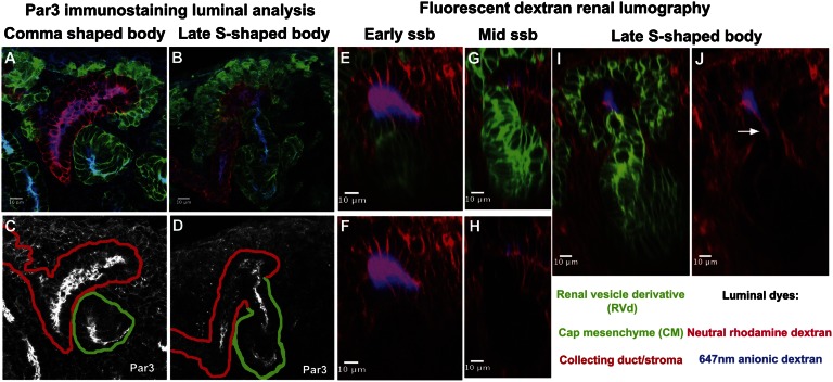
Figure 2.
Luminal interconnection between the nephron and collecting duct systems. (A–J) Stage-specific analysis of connection events using epithelial markers and dye filling; stages and analysis are indicated on the panels. (A–D) Par3 highlights the apical organization of epithelia; continuous labeling between the RV derivative and ureteric epithelium indicates that a continuous epithelium exists at the late S-shaped body stage (compare A and C with B and D). Backfilling with dye injection into the bladder of an intact urogenital system at stage E16.5 shows that dye labels the distal arm of the developing nephron at the late S-shaped body stage (compare E–H with I and J). ssb, S-shaped body.

