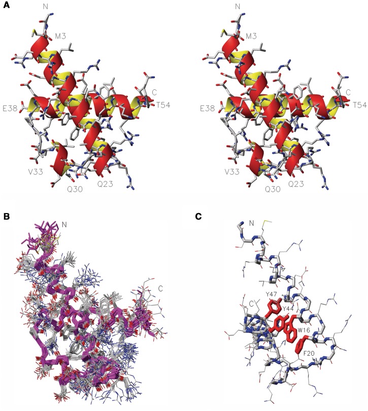
Figure 3.
NMR solution structure of RecQL4_N54. (A) Stereo view (side-by-side) of the structure closest to the mean. Heavy atom colouring: C (grey), N (blue), O (red) and S (yellow). Helices are indicated by an orange/yellow ribbon. Amino acid type and residue numbers for the start and end residue of the helical elements are annotated. (B) Superimposition of the 20 calculated structures with the lowest target function, backbone in magenta, heavy atom colouring as in A. (C) Aromatic core, the side chains of W16, F20, Y44 and Y47 are emphasized in red, aspect rotated with respect to (B) to display the core.

