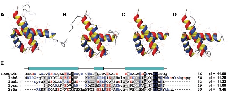
Figure 4.
Superimposition of the NMR solution structure of RecQL4_N54 (orange ribbon) with homeodomains (blue ribbon). (A) Antennapedia homeodomain (PDB code 1AHD; backbone r.m.s.d. 1.87 Å), (B) Engrailed homeodomain (1ENH; 1.86 Å). (C) MATa1/MATα2 homeodomain (1YRN; 1.76 Å). (D) Homeodomain from the SCR/EXD complex (2R5Z; 1.79 Å). Homeodomains in (A), (C) and (D) are in the DNA-bound form, DNA not shown. (E) Structure-based sequence alignment of RecQL4_N54 against the depicted homeodomains. The RecQL4_N54 construct used for structure determination starts with two non-native residues derived from the GST tag after thrombin digest. Numbering of the a.a. positions refers to the construct and not the native RecQL4 sequence to maintain consistency with the related PDB (2KMU) and BMRB (16544) database entries. Therefore, the numbering of the residues is shifted by two compared to the native protein.

