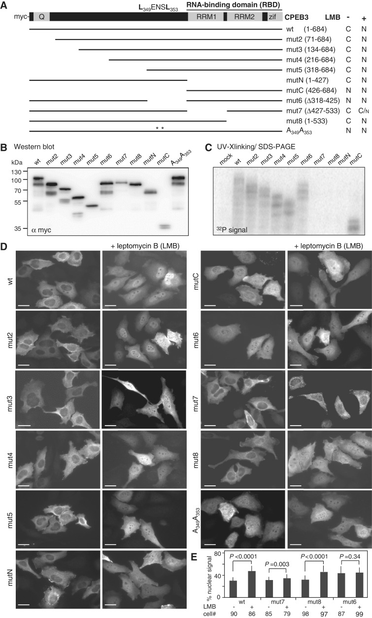
Figure 1.
Identification of the cis-elements required for nucleocytoplasmic translocation of CPEB3. (A) Salient features of CPEB3, showing the N-terminal glutamine-rich region (Q) and the C-terminal RNA-binding domain (RBD) composed of two RNA recognition motifs (RRM) and zinc fingers (Zif). The LENSL motif is the nuclear export sequence. The various myc-tagged CPEB3 mutants are illustrated. The 293 T lysates containing wild-type (wt) and mutant (mut) CPEB3 were (B) used for western blotting or (C) crosslinked with radiolabelled RNA probe for RNA-binding assay. (D) HeLa cells expressing wt and mut CPEB3 were treated with ± 10 ng/ml LMB for 30 min prior to fixation and immunostaining with myc antibody. Subcellular localizations of wt and mut CPEB3 proteins in ± LMB-treated cells are summarized in (A), N: nucleus; C: cytoplasm. Scale: 20 µm (E) To quantify nuclear distribution of mut6, mut7, mut 8 and wt CPEB3, ∼80–100 transfected cells ± LMB treatment were analysed using MetaMorph software. The nuclear CPEB3 signal was divided by total cellular signal to yield the % nuclear signal. Error bars indicate SD. Statistical analysis was performed using Student’s t-test.

