Figure 4.
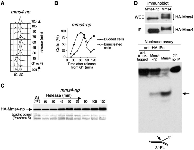
Reduced nuclease activity in a phosphorylation-defective mms4 mutant. (A) HA-mms4-np cells were blocked in G1 using α factor and released from the block in fresh medium. Cells were collected at the indicated time points and the DNA content throughout the cell cycle was monitored using flow cytometry. (B) Percentage of budded and binucleated cells during the experiment. (C) Immunoblot analysis of HA-Mms4-np during the course of the experiment. (D) Nuclease activity assay. The extracts were prepared from HA-mms4-np and HA-MMS4 cells blocked in G2/M with nocodazole. The phosphorylation of wild-type Mms4 and mutant Mms4-np in the whole cell extract (WCE), as well as the yield of the immunoprecipitation of each protein (IP) were monitored by immunoblot (upper panels). About 2% of the total amount of the immunoprecipitated protein used for the nuclease assays was loaded in each case. The nuclease activity was assayed using a 32P-labelled 3′-flap as a substrate (lower panel). An arrow indicates the labelled-product resulting from the nucleolytic cleavage. The controls were as in Figure 3.

