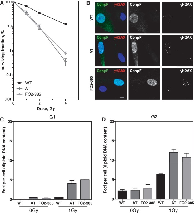
Figure 1.
Similarly enhanced radiosensitivity in AT and Artemis fibroblasts due to an enhanced number of residual DSBs. (A) Radiosensitivity of exponentially growing WT, AT and Artemis cells (FO2-385) was measured by colony formation after various X-ray doses. Error bars represent the SEM of three independent experiments. (B) Differential staining for G2-phase cells (Cenp-F-positive) 24 h after irradiation with 1 Gy in addition to detection of γH2AX foci. Bars, 10 µm. Quantification of residual γH2AX foci in (C) Cenp-F-negative G1-phase cells and (D) Cenp-F-positive G2-phase cells 24 h after IR (1 Gy). S-phase cells showing a strong pan-nuclear γH2AX signal were excluded from the analysis. The AT cell line AT1BR proved to be hyperploid (1.5-fold enhanced compared with WT and Artemis cells (data not shown)). Their foci number/nucleus was normalized to a diploid DNA content.

