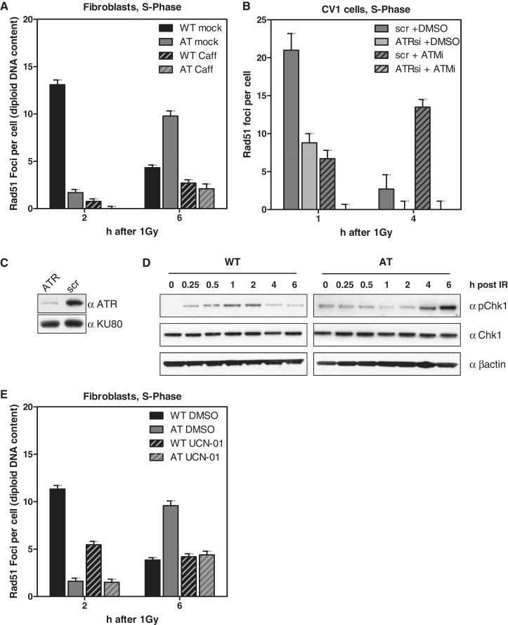
Figure 5.
Rad51 focus formation in AT cells depends on functional ATR. (A) WT and AT cells were pre-treated with 10 mM caffeine for 2 h and pulse-labeled with EdU shortly before IR (1 Gy). Rad51 foci were recorded 2 and 6 h later in EdU-positive cells. Not only the foci number declined upon caffeine treatment but also the size of the remaining foci was reduced (see also Supplementary Figure 5). (B) Quantification of Rad51 foci in EdU-positive CV-1 cells treated with ATR siRNA (C) and scrambled control without ATM inhibition (10 µM KU55933). (D) Western blot of Chk1 expressed in WT and AT cells irradiated with 10 Gy. (E) WT and AT cells were EdU-labeled, irradiated with 1 Gy and during repair continuously exposed to the Chk1 inhibitor UCN-01 (0.1 µM). Quantification of Rad51 foci 2 and 6 h after IR in EdU-positive cells.

