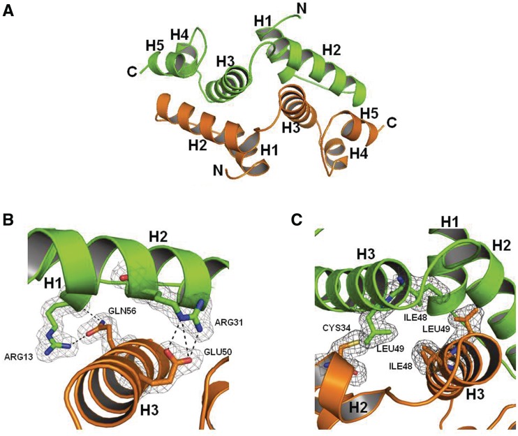
Figure 1.
Overall structure of Zfp206SCAN. (A) The Zfp206SCAN domain-swapped dimer is formed by packing helix H2 of one monomer (green) against helices H3 and helix H5 of the opposing monomer (orange). (B) The (2FO-FC) map of the dimer interface involving residues Arg13 and Arg31 in molecule 1 and Gln56 and Glu50 (as indicated in Figure 2A) in molecule 2 of Zfp206SCAN contacted by hydrogen bond. The electron density (2Fo-Fc) is displayed at the 0.5σ level. (C) The (2FO-FC) map of the dimer interface involving residues Ile48 and Leu49 in molecule 1 and Cys34, Ile48 and Leu49 of molecule 2 (as indicated in Figure 2A).

