Figure 6.
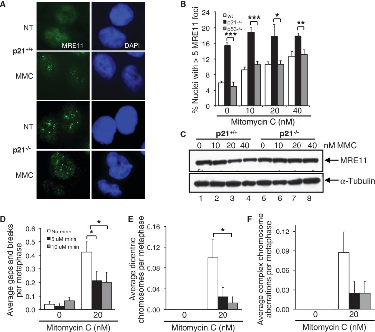
Elevated MRE11 nuclease activity contributes to the increased chromosome instability of p21−/− cells. (A–C) HCT116 wild-type, p21−/− and p53−/− cells were incubated in the absence or presence of 10, 20 and 40 nM MMC for 16 h. (A and B) The number of nuclei displaying >5 discrete MRE11 foci were quantified using immunofluorescence microscopy and plotted. Representative images of MRE11 nuclear foci are shown in (A). At least 300 nuclei were scored per treatment per experiment, and this experiment was performed at least three times with similar results. (C) Whole-cell lysates were prepared and resolved proteins were immunoblotted with anti-MRE11 and anti-α-tubulin antibodies. (D–F) p21−/− cells were incubated in the absence or presence of 20 nM MMC and 5 and 10 µM mirin for 16 h. Metaphase spreads were prepared, and chromosome aberrations, including gaps and breaks (D), dicentric chromosomes (E) and complex aberrations (F), including radial formations, were scored. At least 80 metaphases were scored per treatment, and this experiment was repeated twice with similar findings. Error bars represent standard errors of the means. *P < 0.05; **P < 0.01; ***P < 0.001.

