Figure 4.
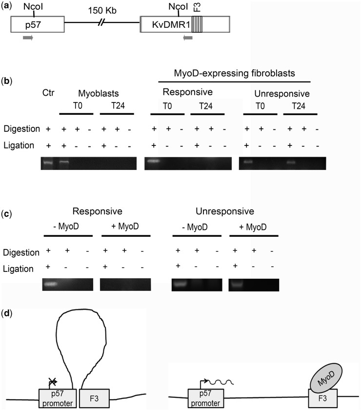
KvDMR1 physically interacts with p57 promoter in undifferentiated and in unresponsive cells. (a) Schematic representation of the p57-KvDMR1 locus showing the locations of the NcoI sites and of the PCR primers (arrows) used for 3C analysis. (b) 3C analysis of the p57-KvDMR1 locus in C2.7 myoblasts and in responsive (C57BL) and in unresponsive (C3H10T1/2) mouse embryo fibroblasts expressing MyoD kept either in growth (T0) or in differentiation medium for 24 h (T24). Ctr consists of a plasmid construct containing a ligation product of p57 promoter and KvDMR1 sequences and represents a positive control for the pair of primers used. The results shown are representative of three independent experiments. (c) 3C analysis of the p57-KvDMR1 locus in responsive and unresponsive fibroblasts infected with either the MyoD retroviral vector (+MyoD) or with the empty vector (–MyoD) and analyzed 24 h after the shift to differentiation medium. The results shown are representative of three independent experiments. (d) Schematic model of MyoD effect on the putative chromatin looping between KvDMR1 and p57 promoter.

