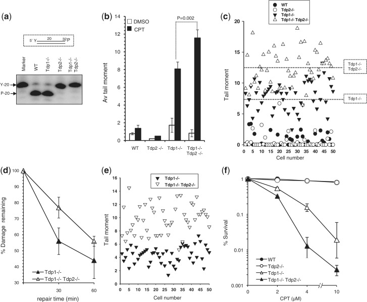
Figure 1.
Murine Tdp2 repairs Top1-mediated DNA damage in the absence of Tdp1. (a) Total cell lysate (10 μg) from WT, Tdp1−/−, Tdp2−/− or Tdp1−/−/Tdp2−/− MEFs was incubated with duplex DNA substrates harbouring the indicated 5′-phosphotyrosine (‘Y’) terminus (inset) and reaction products resolved and detected by denaturing PAGE and phosphorimaging. The positions of oligonucleotide substrate (‘Y-20’) and product (‘P-20’) harbouring 5′-phosphotyrosine and 5′-phosphate termini, respectively, are shown. (b) MEFs of the indicated genotype were incubated with DMSO or 20 μM CPT for 60 min at 37°C and DNA strand breakage quantified by alkaline comet assays. Mean tail moments were quantified for 50 cells/sample/experiment and data are the average of n = 3 biological replicates ± s.e.m. (c) A representative scatter plot of data from one of the experiments in (b) showing comet tail moments of individual cells (50 cells per sample). Dotted lines denote the position of the mean tail moments for the indicated genotypes. (d) MEFs of the indicated genotypes were subjected to CPT treatment as described in (b) followed by subsequent incubation in CPT-free media for a 30- or 60-min repair period. The fraction of DNA breaks remaining was calculated from n = 3 biological replicates and depicted as % damage remaining ± s.e.m. (e) A representative scatter plot of data from the 30 min repair time point from one of the experiments in (d), showing comet tail moments of individual cells (50 cells per sample). (f) MEFs of the indicated genotype were mock-treated or treated with the indicated concentrations of CPT and the number of surviving colonies determined after 7–10 days. Data are from the mean (±s.e.m.) of three independent experiments.

