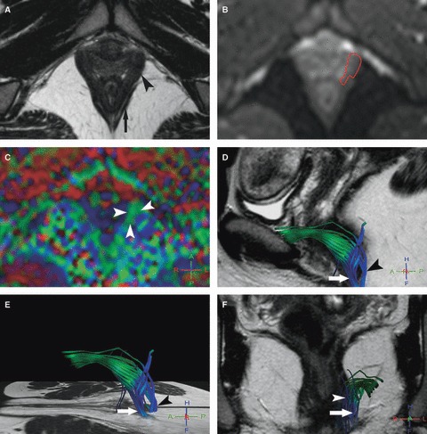
Fig. 1.

Left pubovisceralis MR tractography of a 26-year-old volunteer (score = 3). (A) Axial T2w image; and (B) corresponding axial b0 image show how the manual drawing of the left pubovisceralis (A, arrowhead) was performed on the b0 image (B, red line), excluding the external anal sphincter (A, arrow) with help of the T2w image. (C) The FA map shows a predominantly green color in projection of the pubovisceralis (arrowheads) due to the mainly antero-posterior direction of fibers. (D–F) 3D representations of some fibers in the left pubovisceralis (D, E) in the sagittal and (F) coronal planes with projection on T2w images show accurate green fibers extending posteriorly with a blue encoding in the perineal body (puboperinealis; arrow) and to the lateral canal anal wall (puboanalis; arrowhead). Note also the presence of some blue inaccurate fibers in projection in the ischio-anal adipose tissue.
