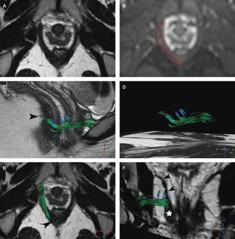
Fig. 2.

Right puborectalis MR tractography of a 22-year-old volunteer (score = 2.5). (A) Axial T2w image; and (B) corresponding axial b0 image show how the manual drawing (B, red line) was performed. (C–F) 3D representation of some fibers in the right puborectalis (C, D) in the sagittal, (E) axial and (F) coronal oblique planes with projection on T2w images show green accurate fibers and the presence of some blue inaccurate fibers. On the coronal view (F) it lies at its characteristic anatomic location lateral and slightly above the pubovisceralis (star), and medial and below the iliococcygeus (arrowheads). Note that fibers around the anorectal junction (C, E, arrowhead) were not tracked.
