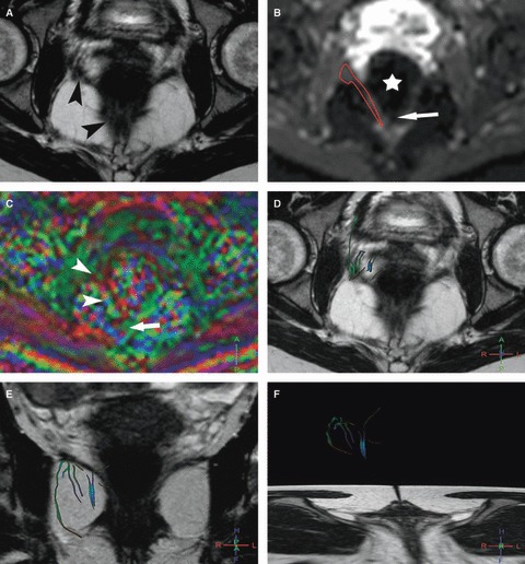
Fig. 3.

Right iliococcygeus MR tractography of a 24-year-old volunteer (score = 0). (A) Axial T2w image; and (B) corresponding axial b0 image show the difficulty of the manual drawing of the right iliococcygeus (arrowheads) particularly at its posterior part with susceptibility artifact (arrow) due to the presence of air within the rectum (star). (C) The FA map shows a random coloration in the area of the iliococcygeus (arrowheads) with distortion artifact at its posterior part (arrow). (D–F) 3D representation of some fibers in the right iliococcygeus (D) in the axial and (E, F) coronal planes with projection on T2w images show sparse and multicolored inaccurate fibers.
