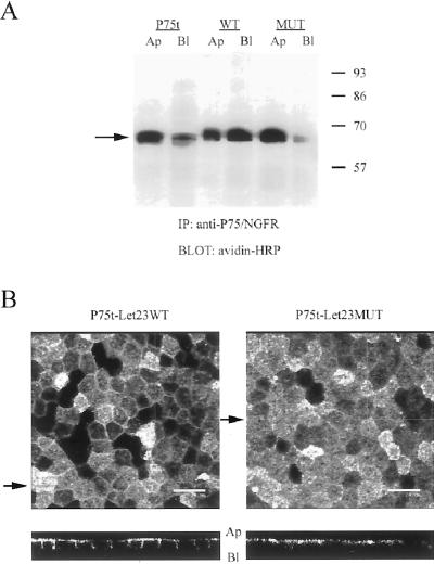
Figure 2.
Differential localization of the receptor chimeras correlated with their ability to bind to mLin-7. Shown in A are the results of a cell-surface biotinylation experiment to determine the steady-state localization of the P75t constructs. Stable MDCK cell lines expressing P75t, P75t-Let23WT, or P75t-Let23MUT were grown as confluent monolayers on Transwell polycarbonate filters and selectively labeled at the apical or basolateral side with biotin. Lysates were collected and immunoprecipitation performed with anti-P75 antibodies. The precipitates were separated by 10% SDS-PAGE, transferred to nitrocellulose, and probed for biotinylated proteins by using avidin-HRP. Relative molecular weight is shown to the left in kilodaltons. An arrow indicates the relevant band. (B) Same cell lines used above were stained with anti-P75 antibodies and secondary antibodies coupled to fluorochromes, and then examined by confocal laser-scanning microscopy. The top panels show digital photomicrographs (the X-Y dimension of the Z-series), whereas the bottom panels show the X-Z dimension (Z-section). The arrows next to the top panels indicate the plane through which the Z-sections were taken. The apical (Ap) and basolateral (Bl) sides are indicated next to the Z-sections. Bars, 20 μm.

