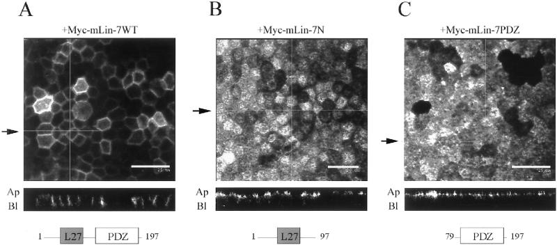
Figure 3.
Myc-mLin-7 deletion mutants interfered with the basolateral localization of P75t-Let23WT. The localization of P75t-Let23WT in MDCK cell lines coexpressing Myc-tagged mLin-7 constructs is shown in A–C: P75t-Let23WT plus Myc-mLin-7WT (full-length mLin-7) (A), Myc-mLin-7N (amino acids 1–92), which contains the mLin-2 binding site (B), or Myc-mLin-7PDZ (amino acids 79–197), containing the PDZ domain (C). These cells were grown as confluent monolayers on Transwell filters, fixed and permeabilized, and then stained with anti-P75 antibodies and secondary antibodies coupled to fluorochromes. Examination was by confocal laser-scanning microscopy. The top panels show digital photomicrographs, whereas the bottom panels show Z-sections. The arrows next to the top panels indicate the plane through which the Z-sections were taken. The apical (Ap) and basolateral (Bl) sides are indicated next to the Z-sections. Bars, 20 (B) and 25 μm (A and C). Below each Z-section is a cartoon depicting the Myc-mLIn-7 construct that was coexpressed in these cell lines. The numbers refer to the amino acid positions, whereas the boxed regions represent identified protein interaction domains: L27, coiled-coil region that binds to mLin-2/CASK; PDZ, PDZ domain that binds to the carboxyl terminus of C. elegans Let-23.

